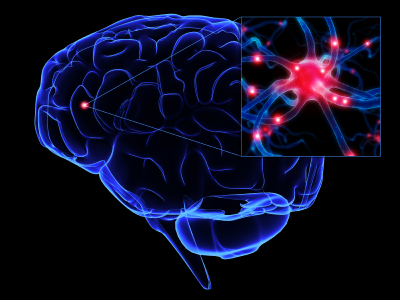
Brain Injuries Under The Microscope
 The steady pace of new information emerging on brain injuries is extremely encouraging.
The latest study, summarized by Reuters, explains how deeper study is detecting brain injury undetected by popular imaging scans.
MAKING ‘THE INVISIBLE INJURY’ VISIBLE
For the new study, published Wednesday in the journal Science Translational Medicine, scientists compared three groups of brains. Four came from military veterans who had suffered the blast of an improvised explosive device (IED) or a concussion. Four belonged to young athletes who had concussions. And scores were from mice that had been exposed to a blast akin to that from an IED 17 feet (five meters) away packed with 12 pounds (5.4 kilograms) of TNT, comparable to an IED made from a 120-mm artillery round.
None of the brains had obvious injury. “If you hold them in your hand you don’t see any damage,” said neuropathologist Ann McKee of the Boston University School of Medicine and the Veterans Affairs New England Healthcare System, co-leader of the new study. “CT and MRI don’t see it. It takes a microscope, even an electron microscope.”
With that scrutiny the damage was clear. Specialized cells called astrocytes extended what BU’s Goldstein called “little feet” that wrapped themselves around blood vessels. Axons crumbled and wound up in cellular garbage cans. Long strings of proteins called tau formed, as seen in Alzheimer’s disease.
We here at the Tampa Bay brain injury blog definitely recommend you read the entire piece.
Google+


